
CREATURE
- HOME
- CREATURE
Examples of analysis
Hepatocytes (pig liver)
x7.000 Inserted images of cell membranes (x100,000)
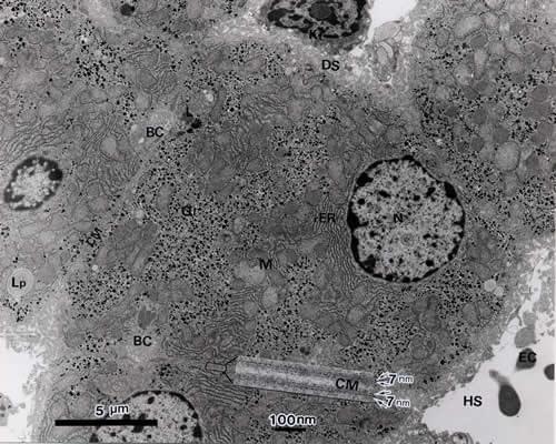
- The liver’s many functions include lipogenesis, protein synthesis, drug metabolism and removal, energy storage, etc. Hepatocytes constitute the bulk of the liver, but bile capillaries (BC) and blood vessels (HS) are also present, and blood cells (EC, Kf) are seen inside blood vessels.
- Each hepatocyte is surrounded by a cell membrane (CM) and has one cell nucleus (N). Cytoplasm is made up of endoplasmic reticulum (rER), which is responsible for protein synthesis, mitochondria (M) for producing cellular energy, and glycogen (Gl) for storing glucose. The inserted image shows the expanded view of cell membranes of adjoining cells, and it shows that each membrane is 7 nm wide and has a double-layer structure.
- The hepatocyte is often used as an example when explaining the structure and functions of a common cell because it contains typical cell organelles.
Wide-angle observation of a glomerulusx1,000 TEM
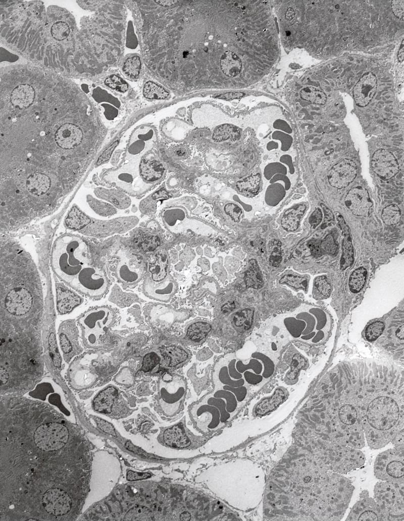
The kidneys are organs that produce urine from blood. A spherical glomerulus, which filters urine from blood, is visible in the center of this image and is surrounded by renal tubules, which reabsorb necessary components from the filtrate.
Pathogenic Escherichia coli O157 x30,000 1:TEM 2:SEM
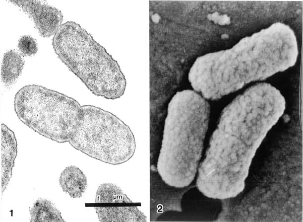
O157 is a bacterium that became known as one of causative bacteria of mass food poisoning in recent years. It is urgently needed to understand its production mechanism of the Vero toxin. Bacteria have cell walls around them, making it difficult for chemicals such as the fixative to penetrate. Although the sample preparation method is basically identical to that for animal tissues, image quality can be enhanced by adding extra process time to steps and performing fixation at room temperature.
Observation of avian adenovirus using the negative staining methodx100,000 TEM TEM
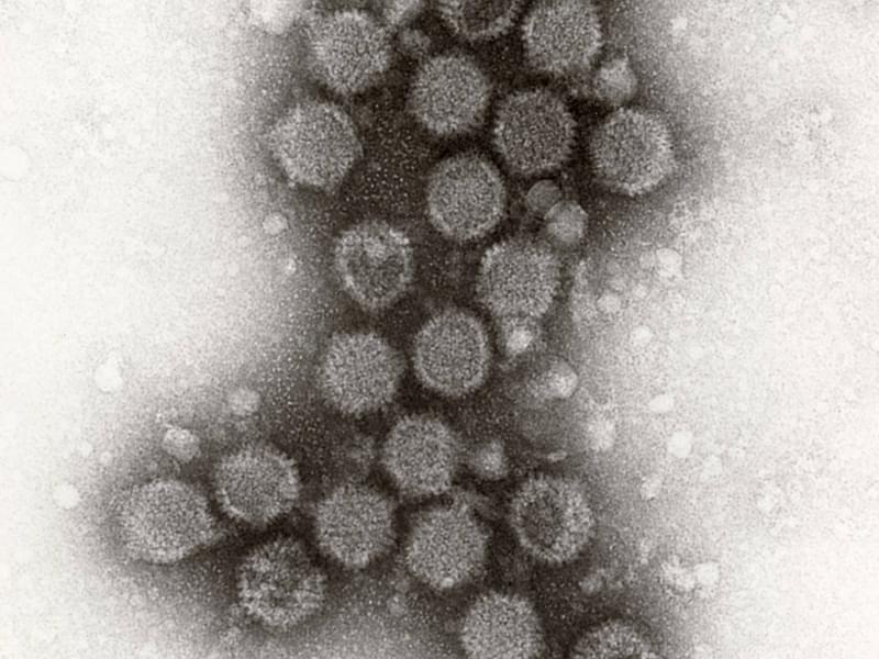
Adenoviruses are said to be major pathogenic viruses for the common cold, and are spherical particles with a diameter of about 80 nm. Spikes of the surface can be observed using the negative staining method.
Mold x2,000
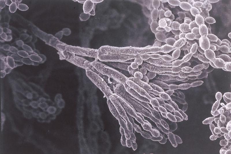
Mold is made up of hyphae and spores. Spores can fall off easily, requiring careful sample preparation work.
Corneal Epithelium (SEM) x1,000
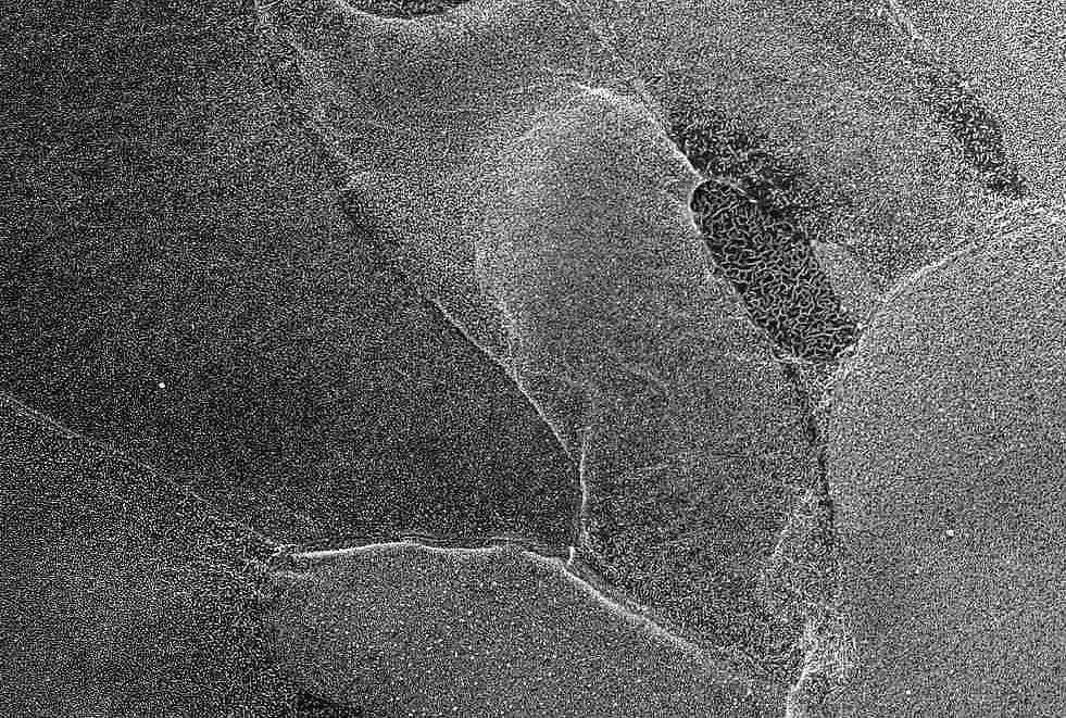
The villi of individual cells can be compared in this image overlooking corneal epithelium cells.
Corneal Epithelium (cross section) x1,500
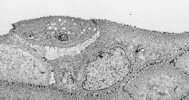
Cross-sectional observation with a TEM allows observation from corneal epithelium cells to the endothelium. High-magnification observation reveals the status of cell adhesion.
Myofibril x2,000
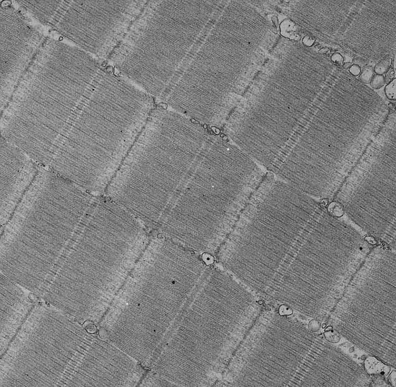
It shows that actin and myosin are lined up neatly with the Z line in the center.
Yeast x1,500
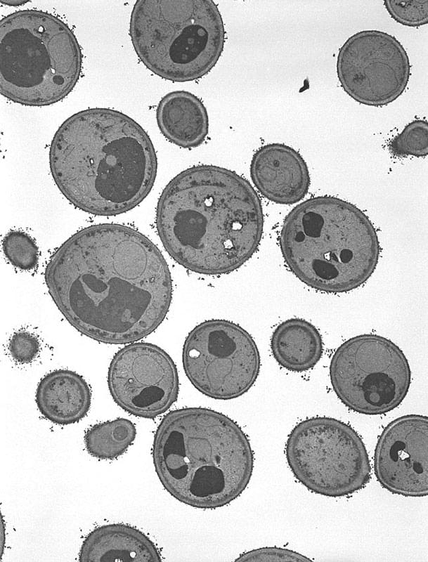
As it is hard to fix yeast, its observation is difficult. There are various organelles inside, and observable organelles may vary depending on the condition of the yeast.
Nerve tissue x3,000
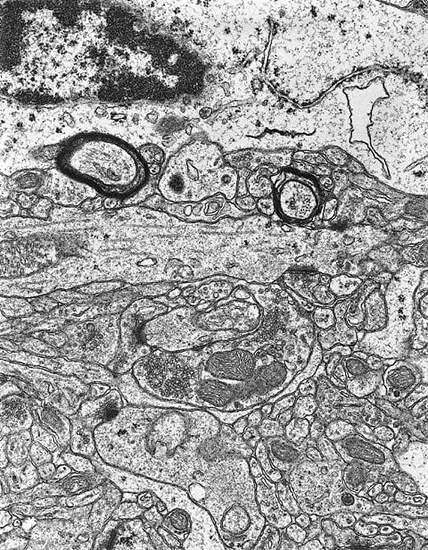
Organelles with unique structures such as myelin structure and synapses exist, and various images can be taken depending of the part.